A | B | C | D | E | F | G | H | CH | I | J | K | L | M | N | O | P | Q | R | S | T | U | V | W | X | Y | Z | 0 | 1 | 2 | 3 | 4 | 5 | 6 | 7 | 8 | 9
| CT scan | |
|---|---|
 Modern CT scanner (2021), photon-counting CT (Siemens NAEOTOM Alpha) | |
| Other names | X-ray computed tomography (X-ray CT), computerized axial tomography scan (CAT scan),[1] computer aided tomography, computed tomography scan |
| ICD-10-PCS | B?2 |
| ICD-9-CM | 88.38 |
| MeSH | D014057 |
| OPS-301 code | 3–20...3–26 |
| MedlinePlus | 003330 |
A computed tomography scan (CT scan; formerly called computed axial tomography scan or CAT scan) is a medical imaging technique used to obtain detailed internal images of the body.[2] The personnel that perform CT scans are called radiographers or radiology technologists.[3][4]
CT scanners use a rotating X-ray tube and a row of detectors placed in a gantry to measure X-ray attenuations by different tissues inside the body. The multiple X-ray measurements taken from different angles are then processed on a computer using tomographic reconstruction algorithms to produce tomographic (cross-sectional) images (virtual "slices") of a body. CT scans can be used in patients with metallic implants or pacemakers, for whom magnetic resonance imaging (MRI) is contraindicated.
Since its development in the 1970s, CT scanning has proven to be a versatile imaging technique. While CT is most prominently used in medical diagnosis, it can also be used to form images of non-living objects. The 1979 Nobel Prize in Physiology or Medicine was awarded jointly to South African-American physicist Allan MacLeod Cormack and British electrical engineer Godfrey Hounsfield "for the development of computer-assisted tomography".[5][6]
Types
On the basis of image acquisition and procedures, various type of scanners are available in the market.
Sequential CT
Sequential CT, also known as step-and-shoot CT, is a type of scanning method in which the CT table moves stepwise. The table increments to a particular location and then stops which is followed by the X-ray tube rotation and acquisition of a slice. The table then increments again, and another slice is taken. The table movement stops while taking slices. This results in an increased time of scanning.[7]
Spiral CT
_and_patient_in_a_CT_imaging_system.gif)

Spinning tube, commonly called spiral CT, or helical CT, is an imaging technique in which an entire X-ray tube is spun around the central axis of the area being scanned. These are the dominant type of scanners on the market because they have been manufactured longer and offer a lower cost of production and purchase. The main limitation of this type of CT is the bulk and inertia of the equipment (X-ray tube assembly and detector array on the opposite side of the circle) which limits the speed at which the equipment can spin. Some designs use two X-ray sources and detector arrays offset by an angle, as a technique to improve temporal resolution.[8][9]
Electron beam tomography
Electron beam tomography (EBT) is a specific form of CT in which a large enough X-ray tube is constructed so that only the path of the electrons, travelling between the cathode and anode of the X-ray tube, are spun using deflection coils.[10] This type had a major advantage since sweep speeds can be much faster, allowing for less blurry imaging of moving structures, such as the heart and arteries.[11] Fewer scanners of this design have been produced when compared with spinning tube types, mainly due to the higher cost associated with building a much larger X-ray tube and detector array and limited anatomical coverage.[12]
Dual Energy CT
Dual Energy CT, also known as Spectral CT, is an advancement of Computed Tomography in which two energies are used to create two sets of data.[13] A Dual Energy CT may employ Dual source, Single source with dual detector layer, Single source with energy switching methods to get two different sets of data.[14]
- Dual source CT is an advanced scanner with a two X-ray tube detector system, unlike conventional single tube systems.[15][16] These two detector systems are mounted on a single gantry at 90° in the same plane.[17] Dual Source CT scanners allow fast scanning with higher temporal resolution by acquiring a full CT slice in only half a rotation. Fast imaging reduces motion blurring at high heart rates and potentially allowing for shorter breath-hold time. This is particularly useful for ill patients having difficulty holding their breath or unable to take heart-rate lowering medication.[17][18]
- Single Source with Energy switching is another mode of Dual energy CT in which a single tube is operated at two different energies by switching the energies frequently.[19][20]
CT perfusion imaging

CT perfusion imaging is a specific form of CT to assess flow through blood vessels whilst injecting a contrast agent.[21] Blood flow, blood transit time, and organ blood volume, can all be calculated with reasonable sensitivity and specificity.[21] This type of CT may be used on the heart, although sensitivity and specificity for detecting abnormalities are still lower than for other forms of CT.[22] This may also be used on the brain, where CT perfusion imaging can often detect poor brain perfusion well before it is detected using a conventional spiral CT scan.[21][23] This is better for stroke diagnosis than other CT types.[23]
PET CT

Positron emission tomography–computed tomography is a hybrid CT modality which combines, in a single gantry, a positron emission tomography (PET) scanner and an X-ray computed tomography (CT) scanner, to acquire sequential images from both devices in the same session, which are combined into a single superposed (co-registered) image. Thus, functional imaging obtained by PET, which depicts the spatial distribution of metabolic or biochemical activity in the body can be more precisely aligned or correlated with anatomic imaging obtained by CT scanning.[24]
PET-CT gives both anatomical and functional details of an organ under examination and is helpful in detecting different type of cancers.[25][26]
Medical use
Since its introduction in the 1970s,[27] CT has become an important tool in medical imaging to supplement conventional X-ray imaging and medical ultrasonography. It has more recently been used for preventive medicine or screening for disease, for example, CT colonography for people with a high risk of colon cancer, or full-motion heart scans for people with a high risk of heart disease. Several institutions offer full-body scans for the general population although this practice goes against the advice and official position of many professional organizations in the field primarily due to the radiation dose applied.[28]
The use of CT scans has increased dramatically over the last two decades in many countries.[29] An estimated 72 million scans were performed in the United States in 2007 and more than 80 million in 2015.[30][31]
Head

CT scanning of the head is typically used to detect infarction (stroke), tumors, calcifications, haemorrhage, and bone trauma.[32] Of the above, hypodense (dark) structures can indicate edema and infarction, hyperdense (bright) structures indicate calcifications and haemorrhage and bone trauma can be seen as disjunction in bone windows. Tumors can be detected by the swelling and anatomical distortion they cause, or by surrounding edema. CT scanning of the head is also used in CT-guided stereotactic surgery and radiosurgery for treatment of intracranial tumors, arteriovenous malformations, and other surgically treatable conditions using a device known as the N-localizer.[33][34][35][36][37][38]
Neck
Contrast CT is generally the initial study of choice for neck masses in adults.[39] CT of the thyroid plays an important role in the evaluation of thyroid cancer.[40] CT scan often incidentally finds thyroid abnormalities, and so is often the preferred investigation modality for thyroid abnormalities.[40]
Lungs
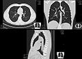
A CT scan can be used for detecting both acute and chronic changes in the lung parenchyma, the tissue of the lungs.[41] It is particularly relevant here because normal two-dimensional X-rays do not show such defects. A variety of techniques are used, depending on the suspected abnormality. For evaluation of chronic interstitial processes such as emphysema, and fibrosis,[42] thin sections with high spatial frequency reconstructions are used; often scans are performed both on inspiration and expiration. This special technique is called high resolution CT that produces a sampling of the lung, and not continuous images.[43]
_and_diameter_(D).svg)
Bronchial wall thickening can be seen on lung CTs and generally (but not always) implies inflammation of the bronchi.[44]
An incidentally found nodule in the absence of symptoms (sometimes referred to as an incidentaloma) may raise concerns that it might represent a tumor, either benign or malignant.[45] Perhaps persuaded by fear, patients and doctors sometimes agree to an intensive schedule of CT scans, sometimes up to every three months and beyond the recommended guidelines, in an attempt to do surveillance on the nodules.[46] However, established guidelines advise that patients without a prior history of cancer and whose solid nodules have not grown over a two-year period are unlikely to have any malignant cancer.[46] For this reason, and because no research provides supporting evidence that intensive surveillance gives better outcomes, and because of risks associated with having CT scans, patients should not receive CT screening in excess of those recommended by established guidelines.[46]
Angiography

Computed tomography angiography (CTA) is a type of contrast CT to visualize the arteries and veins throughout the body.[47] This ranges from arteries serving the brain to those bringing blood to the lungs, kidneys, arms and legs. An example of this type of exam is CT pulmonary angiogram (CTPA) used to diagnose pulmonary embolism (PE). It employs computed tomography and an iodine-based contrast agent to obtain an image of the pulmonary arteries.[48][49][50]
Cardiac
A CT scan of the heart is performed to gain knowledge about cardiac or coronary anatomy.[51] Traditionally, cardiac CT scans are used to detect, diagnose, or follow up coronary artery disease.[52] More recently CT has played a key role in the fast-evolving field of transcatheter structural heart interventions, more specifically in the transcatheter repair and replacement of heart valves.[53][54][55]
The main forms of cardiac CT scanning are:
- Coronary CT angiography (CCTA): the use of CT to assess the coronary arteries of the heart. The subject receives an intravenous injection of radiocontrast, and then the heart is scanned using a high-speed CT scanner, allowing radiologists to assess the extent of occlusion in the coronary arteries, usually to diagnose coronary artery disease.[56][57]
- Coronary CT calcium scan: also used for the assessment of severity of coronary artery disease. Specifically, it looks for calcium deposits in the coronary arteries that can narrow arteries and increase the risk of a heart attack.[58] A typical coronary CT calcium scan is done without the use of radiocontrast, but it can possibly be done from contrast-enhanced images as well.[59]
To better visualize the anatomy, post-processing of the images is common.[52] Most common are multiplanar reconstructions (MPR) and volume rendering. For more complex anatomies and procedures, such as heart valve interventions, a true 3D reconstruction or a 3D print is created based on these CT images to gain a deeper understanding.[60][61][62][63]
Abdomen and pelvis

CT is an accurate technique for diagnosis of abdominal diseases like Crohn's disease,[64] GIT bleeding, and diagnosis and staging of cancer, as well as follow-up after cancer treatment to assess response.[65] It is commonly used to investigate acute abdominal pain.[66]
Non-enhanced computed tomography is today the gold standard for diagnosing urinary stones.[67] The size, volume and density of stones can be estimated to help clinicians guide further treatment; size is especially important in predicting spontaneous passage of a stone.[68]
Axial skeleton and extremities
For the axial skeleton and extremities, CT is often used to image complex fractures, especially ones around joints, because of its ability to reconstruct the area of interest in multiple planes. Fractures, ligamentous injuries, and dislocations can easily be recognized with a 0.2 mm resolution.[69][70] With modern dual-energy CT scanners, new areas of use have been established, such as aiding in the diagnosis of gout.[71]
Biomechanical use
CT is used in biomechanics to quickly reveal the geometry, anatomy, density and elastic moduli of biological tissues.[72][73]
Other uses
Industrial use
Industrial CT scanning (industrial computed tomography) is a process which uses X-ray equipment to produce 3D representations of components both externally and internally. Industrial CT scanning has been used in many areas of industry for internal inspection of components. Some of the key uses for CT scanning have been flaw detection, failure analysis, metrology, assembly analysis, image-based finite element methods[74] and reverse engineering applications. CT scanning is also employed in the imaging and conservation of museum artifacts.[75]
Aviation security
CT scanning has also found an application in transport security (predominantly airport security) where it is currently used in a materials analysis context for explosives detection CTX (explosive-detection device)[76][77][78][79] and is also under consideration for automated baggage/parcel security scanning using computer vision based object recognition algorithms that target the detection of specific threat items based on 3D appearance (e.g. guns, knives, liquid containers).[80][81][82] Its usage in airport security pioneered at Shannon Airport in March 2022 has ended the ban on liquids over 100 ml there, a move that Heathrow Airport plans for a full roll-out on 1 December 2022 and the TSA spent $781.2 million on an order for over 1,000 scanners, ready to go live in the summer.
Geological use
X-ray CT is used in geological studies to quickly reveal materials inside a drill core.[83] Dense minerals such as pyrite and barite appear brighter and less dense components such as clay appear dull in CT images.[84]
Cultural heritage use
X-ray CT and micro-CT can also be used for the conservation and preservation of objects of cultural heritage. For many fragile objects, direct research and observation can be damaging and can degrade the object over time. Using CT scans, conservators and researchers are able to determine the material composition of the objects they are exploring, such as the position of ink along the layers of a scroll, without any additional harm. These scans have been optimal for research focused on the workings of the Antikythera mechanism or the text hidden inside the charred outer layers of the En-Gedi Scroll. However, they are not optimal for every object subject to these kinds of research questions, as there are certain artifacts like the Herculaneum papyri in which the material composition has very little variation along the inside of the object. After scanning these objects, computational methods can be employed to examine the insides of these objects, as was the case with the virtual unwrapping of the En-Gedi scroll and the Herculaneum papyri.[85] Micro-CT has also proved useful for analyzing more recent artifacts such as still-sealed historic correspondence that employed the technique of letterlocking (complex folding and cuts) that provided a "tamper-evident locking mechanism".[86][87] Further examples of use cases in archaeology is imaging the contents of sarcophagi or ceramics.[88]
Recently, CWI in Amsterdam has collaborated with Rijksmuseum to investigate art object inside details in the framework called IntACT.[89]
Micro organism research
Varied types of fungus can degrade wood to different degrees, one Belgium research group has been used X-ray CT 3 dimension with sub-micron resolution unveiled fungi can penetrate micropores of 0.6 μm[90] under certain conditions.
Timber sawmill
Sawmills use industrial CT scanners to detect round defects, for instance knots, to improve total value of timber productions. Most sawmills are planning to incorporate this robust detection tool to improve productivity in the long run, however initial investment cost is high.
Interpretation of resultsedit
Presentationedit

− Average intensity projection
− Maximum intensity projection
− Thin slice (median plane)
− Volume rendering by high and low threshold for radiodensity
The result of a CT scan is a volume of voxels, which may be presented to a human observer by various methods, which broadly fit into the following categories:
- Slices (of varying thickness). Thin slice is generally regarded as planes representing a thickness of less than 3 mm.[91][92] Thick slice is generally regarded as planes representing a thickness between 3 mm and 5 mm.[92][93]
- Projection, including maximum intensity projection[94] and average intensity projection
- Volume rendering (VR)[94]
Technically, all volume renderings become projections when viewed on a 2-dimensional display, making the distinction between projections and volume renderings a bit vague. The epitomes of volume rendering models feature a mix of for example coloring and shading in order to create realistic and observable representations.[95][96]
Two-dimensional CT images are conventionally rendered so that the view is as though looking up at it from the patient's feet.[97] Hence, the left side of the image is to the patient's right and vice versa, while anterior in the image also is the patient's anterior and vice versa. This left-right interchange corresponds to the view that physicians generally have in reality when positioned in front of patients.[98]
Grayscaleedit
Pixels in an image obtained by CT scanning are displayed in terms of relative radiodensity. The pixel itself is displayed according to the mean attenuation of the tissue(s) that it corresponds to on a scale from +3,071 (most attenuating) to −1,024 (least attenuating) on the Hounsfield scale. A pixel is a two dimensional unit based on the matrix size and the field of view. When the CT slice thickness is also factored in, the unit is known as a voxel, which is a three-dimensional unit.[99] Water has an attenuation of 0 Hounsfield units (HU), while air is −1,000 HU, cancellous bone is typically +400 HU, and cranial bone can reach 2,000 HU.[100] The attenuation of metallic implants depends on the atomic number of the element used: Titanium usually has an amount of +1000 HU, iron steel can completely block the X-ray and is, therefore, responsible for well-known line-artifacts in computed tomograms. Artifacts are caused by abrupt transitions between low- and high-density materials, which results in data values that exceed the dynamic range of the processing electronics.[101]
Windowingedit
CT data sets have a very high dynamic range which must be reduced for display or printing. This is typically done via a process of "windowing", which maps a range (the "window") of pixel values to a grayscale ramp. For example, CT images of the brain are commonly viewed with a window extending from 0 HU to 80 HU. Pixel values of 0 and lower, are displayed as black; values of 80 and higher are displayed as white; values within the window are displayed as a gray intensity proportional to position within the window.[102] The window used for display must be matched to the X-ray density of the object of interest, in order to optimize the visible detail.[103] Window width and window level parameters are used to control the windowing of a scan.[104]
Multiplanar reconstruction and projectionsedit


Multiplanar reconstruction (MPR) is the process of converting data from one anatomical plane (usually transverse) to other planes. It can be used for thin slices as well as projections. Multiplanar reconstruction is possible as present CT scanners provide almost isotropic resolution.[105]
MPR is used almost in every scan. The spine is frequently examined with it.[106] An image of the spine in axial plane can only show one vertebral bone at a time and cannot show its relation with other vertebral bones. By reformatting the data in other planes, visualization of the relative position can be achieved in sagittal and coronal plane.[107]
New software allows the reconstruction of data in non-orthogonal (oblique) planes, which help in the visualization of organs which are not in orthogonal planes.[108][109] It is better suited for visualization of the anatomical structure of the bronchi as they do not lie orthogonal to the direction of the scan.[110]
Curved-plane reconstruction (or curved planar reformation = CPR) is performed mainly for the evaluation of vessels. This type of reconstruction helps to straighten the bends in a vessel, thereby helping to visualize a whole vessel in a single image or in multiple images. After a vessel has been "straightened", measurements such as cross-sectional area and length can be made. This is helpful in preoperative assessment of a surgical procedure.[111]
For 2D projections used in radiation therapy for quality assurance and planning of external beam radiotherapy, including digitally reconstructed radiographs, see Beam's eye view.
| Type of projection | Schematic illustration | Examples (10 mm slabs) | Description | Uses |
|---|---|---|---|---|
| Average intensity projection (AIP) | 
|
The average attenuation of each voxel is displayed. The image will get smoother as slice thickness increases. It will look more and more similar to conventional projectional radiography as slice thickness increases. | Zdroj:https://en.wikipedia.org?pojem=CT_scan