A | B | C | D | E | F | G | H | CH | I | J | K | L | M | N | O | P | Q | R | S | T | U | V | W | X | Y | Z | 0 | 1 | 2 | 3 | 4 | 5 | 6 | 7 | 8 | 9
| Anterior cardinal vein | |
|---|---|
 Scheme of arrangement of parietal veins | |
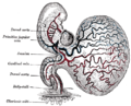
 Human embryo with heart and anterior body-wall removed to show the sinus venosus and its tributaries. | |
| Details | |
| Carnegie stage | 13 |
| Gives rise to | Internal jugular veins and Superior vena cava |
| System | Cardiovascular system |
| Identifiers | |
| Latin | Venae precardinalis |
| TE | cardinal vein_by_E5.11.2.2.2.2.2 E5.11.2.2.2.2.2 |
| Anatomical terminology | |
The anterior cardinal veins (precardinal veins) contribute to the formation of the internal jugular veins and together with the common cardinal vein form the superior vena cava.
The anastomosis between the two anterior cardinal veins develops into the left brachiocephalic vein.[1]
Additional images
-
Human embryo of about fourteen days, with yolk sac.

See also
References
![]() This article incorporates text in the public domain from page 520 of the 20th edition of Gray's Anatomy (1918)
This article incorporates text in the public domain from page 520 of the 20th edition of Gray's Anatomy (1918)
- ^ Sadler, T. W. (2019). Langman's medical embryology (Fourteenth ed.). Philadelphia. ISBN 978-1-4963-8390-7. OCLC 1042400100.
{{cite book}}: CS1 maint: location missing publisher (link)
External links
This cardiovascular system article is a stub. You can help Wikipedia by expanding it. |
>Text je dostupný pod licencí Creative Commons Uveďte autora – Zachovejte licenci, případně za dalších podmínek. Podrobnosti naleznete na stránce Podmínky užití.
Text je dostupný za podmienok Creative
Commons Attribution/Share-Alike License 3.0 Unported; prípadne za ďalších
podmienok.
Podrobnejšie informácie nájdete na stránke Podmienky
použitia.